Ever wondered how your dog’s brain works? A brief guide…
Your dog’s brain is a sophisticated organ – it controls his thinking, learning, and actions. It’s also responsible for interpreting and integrating information from all over the body, much like our human brains. And, in February 2014, research led by Dr Attila Andics revealed more similarities.
The study made by the University of Budapest revealed that a dog’s brain reacts to voices in the same way as a human brain. Eleven dogs and owners where each placed in an MRI scanner and played over 200 different sounds – from car sounds and whistles to human sounds and dog noises. The researchers found that a similar region – the temporal pole, which is the most anterior part of the temporal lobe, was activated when both the animals and people heard human voices. “We do know there are voice areas in humans, areas that respond more strongly to human sounds that any other types of sounds,” Dr Andics explained. “The location (of the activity) in the dog brain is very similar to where we found it in the human brain. The fact that we found these areas exist at all in the dog brain at all is a surprise – it is the first time we have seen this in a non-primate.” The fact that emotionally charged sounds, such as crying or laughter prompted similar responses to humans as it did with the dogs tested might also perhaps explain why dogs are attuned to human emotions.
But what else can we learn by studying our pet’s brain? Here’s a brief guide….
The size and weight of the brain varies greatly from species; the weight of the brain in an average dog is less than half of one per cent of its body weight – but it receives over twenty per cent of the blood pumped out of the heart. So, this shows how the brain is at the core of your dog’s activity, busy digesting data and determining the best course of action, which affects your pooch’s overall behaviour.
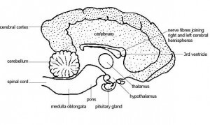
The brain is a mass of nerve tissue which is divided up into three main areas; the cerebrum, the cerebellum and the brain stem. Each part performs particular functions with information being fed into these key areas, so collectively they give instructions on the appropriate action.
The cerebrum or cerebral cortex forms the bulk of the brain. This is responsible for receiving and analysing sensory information such as vision, hearing, touch, taste and pain. The larger the cerebral cortex in an animal, the more options of responses it has, enabling it to carry out complex behaviour patterns. For example; reptiles’ cerebral cortex is far less developed compared to your dog’s brain. This means, Fido can perform many tasks and has complex behaviour patterns compared to the reptile.
The cerebral cortex is divided up into two areas; the left and right cerebral hemispheres. The narrow slit separating these hemispheres is called the cerebral longitudinal fissure. Within these two areas are four lobes; the frontal, temporal, parietal and occipital lobe. The frontal and temporal lobes contribute to the alertness, intelligence (planning and execution of movements), memory and temperament of the dog. Within this area is the thalamus. This is responsible for relaying sensory information such as hearing, sight, touch and pain. The thalamus also enables your dog to selectively concentrate and focus on one thing at a time. The sensory and emotional information relayed to the thalamus is then sent to the parietal and occipital lobes of the dog’s brain for decoding. Once this information has been digested and processed according to previous experiences or memories, the data is then sent to the frontal lobe and translated into plans and actions. The thalamus also contributes to the monitoring and regulation of motor activity initiated in the cerebral cortex. This information is then sent from the cerebral cortex to the cerebellum to aid the co-coordinating centre of the brain which is responsible for muscle activity.
Just below the thalamus is the hypothalamus. This area controls the release of the pituitary hormones (from the pituitary gland) and is responsible for regulating your pet’s drinking and eating behaviour, as well as his body temperature, reproductive and autonomic nervous system; this system contains nerves which control involuntary movements of organs such as the intestines, heart, blood vessels and blood (dogs do not have voluntary control over the autonomic nervous system). Interestingly, emotions such as rage and aggression originate in the hypothalamus – although these are normally inhibited by the hippocampus and the frontal lobe of the cerebral cortex – if a dog contracts the rabies virus, this invades the hippocampus and removes this inhibition. This means the powerful aggressive urges of the hypothalamus are allowed to prevail. As you can see, your dog’s brain is a complex machine, and within the cerebral cortex is the limbic system – this regulates the dog’s emotions from fear, rage, and aggression to anxiety, joy and euphoria. It has an essential role in the learning process. The rabies virus will attack the limbic system and this demonstrates how any disturbances in this area can cause emotional and or behavioural problems.
Within the limbic system is the amygdala, this is responsible for survival strategies and defense responses. In times of extreme danger or a life and death situation a dog has to act quickly. So, in this instance the information of this situation is sent directly from the thalamus to the amygdala activating your dog’s defence reactions at speed, rather than it being decoded first by the cerebral cortex which takes longer to process.
Little brain…
The second area of your dog’s brain is the cerebellum (meaning ‘little brain’ in Latin). This is located at the back of the brain and is attached to the brain stem and cerebral cortex. The cerebellum is the part of the brain that regulates or is mostly responsible for the control and co-ordination of voluntary movement (muscles) and posture of your dog. The cerebellum is interconnected via thalamic relays with the sensory-motor area of the cerebral cortex. So, the cerebellum will receive information from the cerebral cortex about intended muscle activity and it will process and compare this information from receptors in your dog’s muscles and tendons. Once the cerebellum has feedback the data, this ensures precision in movement. Any damage or cerebellar lesioning to this area will typically cause head or body tremors, poor balance, signs of clumsiness, exaggerated and awkward movements. The cerebellum, which is responsible for co-ordinated movement and the rest of the nervous system, is not fully developed at birth. While the brain, spinal cord and associated nerves are all present, the nerves lack the ability to efficiently transmit electrical impulses. Most people who have seen a new born puppy will notice how they are sluggish in movement and its pain sensation is very slow.
Brain stem
The third area of the brain is the brain stem. This is located at the base of the brain and is connected to the spinal cord and cerebellum. There are two main parts of the brain stem; the pons and the medulla oblongata. All the nerve fibres leaving the brain going to your dog’s muscles, will pass through the brain stem. The medulla oblongata is situated at the base of the brain and connects to the spinal cord. It’s responsible for regulating a number of functions from your dog’s heart beat and his breathing to salivation, coughing, sneezing and his gastrointestinal functions. The medulla oblongata, together with the pons, is an important relay site for hearing and balance information, taste sensations and motor reactions. The pons provides a pathway for the nerves fibres to relay sensory information between the cerebellum and cerebral cortex. The pons also includes the micturition centre (urination). Studies in 1964 by Japanese scientists Kuru and Yamamoto, demonstrated how electrical stimulation to the pons resulted in an increase in urethral sphincter activity and the relaxation of the bladder. So, it’s safe to assume that damage to the pons will contribute to urinary incontinence.
How does the brain receive and transmit information?
The central nervous system is comprised of the brain and spinal cord, but connected to these are a network of peripheral nerves (the peripheralnervous system) which penetrate and supply the tissues of the body and transmit pieces of information – such as pain sensation – to and from the body back to the nervous system. In turn, the brain reacts with a course of action. The brain cells that transmit information within the central nervous system are called neurones. Structurally a neuron is unlike any other cell in the body, made up of three parts; the cell body, an axon and dendrites.
The cell body is the large central portion of the cell containing the nucleus and is between the axon and dendrites. The axon is a slender tube that carries nerve impulses away from the neuron to the terminal buttons. Dendrites are short and tree-like; they receive messages from the other neurons. Between the axon terminal button of one cell (presynaptic cell) and the dendrites of the second or receiving cell (postsynaptic cell) is a junction called the synapse. The axon and dendrite in the two cells face each other and the synapse is the very small gap in between. When the information (referred to as the action potential) is being sent through the neurons, the axon terminal of the sending cell triggers the release of a chemical (neurotransmitter) in the immediate area of the dendrite of the receiving cell. Chemicals secreted include; dopamine, noradrenalin and serotonin. And, it is these three neurotransmitters that are important in the treatment of canine behaviour problems. That’s because neurotransmitters can excite, inhibit or alter the activity of other neurons.
Trainer Val Strong uses an analogy that helps us understand how neurotransmitters can excite or inhibit cells. She refers to the receptors on the receiving cell’s membrane like ‘locked doors’. Excitatory neurotransmitters act like keys which open the ‘doors’ allowing information to be passed along the axon of the cell, causing the release of the second cell’s chemical (or neurotransmitter). Whereas inhibitory neurotransmitters acts as ‘bolts’, bolting the receptor doors so the action from the excitatory transmitters have no effect. Changes to the responses of synapses are believed to be the key to memory and learning.
Phew!
As you can see, your dog’s brain is a powerful organ that’s super sophisicated – enabling him to learn, express emotions and allows for behaviour – helping him to respond and adapt to his environment. So, there’s an awful lot going on ‘up top’!
Learn more about our classes

Get Hanne's Book
Playing With Your Dog will help any dog owner work out the games that are best suited for their pet to play throughout his life, from puppyhood to old age. The book also shares some tricks for all ages, group activities, and recommended toys that dogs will enjoy.


